|
Click
on the scans for more detailed views
|
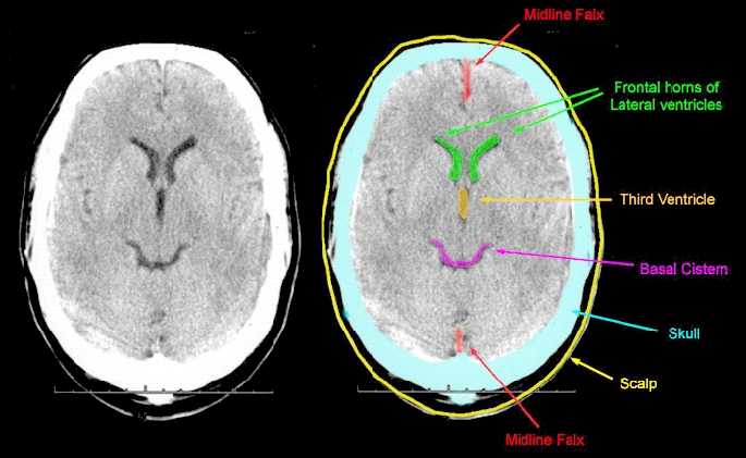
| Case 1 | Normal Scan |
 |
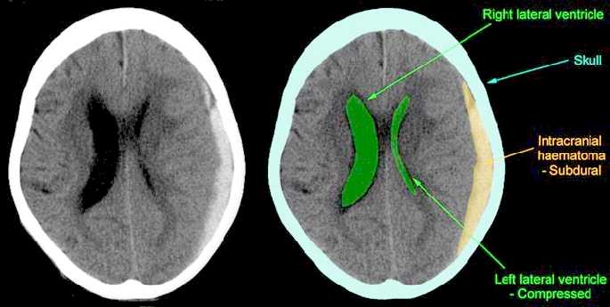
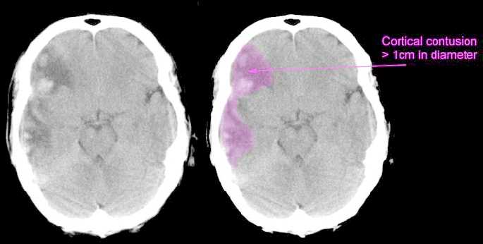
| Case 2 | Acute
subdural haematoma demonstrating midline shift Midline shift > 5mm Intracranial haematoma - non-evacuated Cortical contusion > 1cm in diameter Obliteration of the 3rd ventricle |
 |
| Case 3 | Acute
subdural haematoma Intracranial haematoma - non-evacuated |
 |
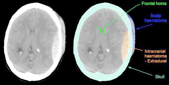
| Case 4 | Acute
extradural haematoma Intracranial haematoma - non-evacuated |
 |
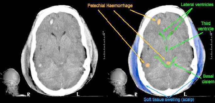
| Case 5 | Diffuse
axonal injury One or more petechial haemorrhages within the brain |
 |
| Case 6 | Cerebral
contusion Cortical contusion > 1cm in diameter |
 |
| Early Outcome Form Section 6 Head CT scan - subheadings |
| Obliteration of the third ventricle |
The third ventricle is demonstrated well in Case One - Normal Scan. Here you can see that it is effectively a small cleft within the brain. If there is any pressure, or swelling of the brain, this is one of the first structures to disappear on the scan, as the walls of the cleft are pushed against each other. It is a sign of increased pressure within the head and can either result from a blood clot pressing on the brain, or of swelling of the brain itself. |
| Midline shift - Greater than 5mm |
If you look at Case Two, you will see that we have drawn the midline using a series of yellow dots. You can do the same thing by using a ruler and joining the falx cerebri anteriorly and posteriorly, as labelled in Case One. The third ventricle and the septum between the frontal horns of the lateral ventricle should not deviate from the midline at all, although there may be slight variations less than 5mm. You can use the scale to the right hand side of each scan to work out if the shift is greater than 5mm. |
| Intracranial haematoma - Evacuated | Often, when you look at a post operative scan, there is still residual blood clot. Even though an extradural, or subdural haematoma has been removed, one will often see evidence of a blood clot post operatively, which is hopefully much smaller and not causing pressure on the brain. It does, however, indicate that there has been evacuation of a significant intracranial haematoma. These cases should be classified as "Intracranial haematoma - Evacuated" |
|
Intracranial
haematoma - |
This refers to blood clot lying either on the surface of the brain in the extradural, or subdural space, or, indeed, to haematomas within the brain substance itself. If these have not been removed surgically, then they should be classified as non-evacuated. |
| One, or more petechial haemorrhages within the brain. |
This refers to very small haemorrhages seen as small, white dots on the scan. They usually occur at the interface between grey and white matter (See Case Five). Other classical sites are the dorsolateral quadrant of the midbrain and also the corpus callosum. They are an indicator of diffuse axonal injury, which is a form of severe primary injury to the brain. This usually carries quite a poor prognosis. |