Back

Case 1 - Normal CT Scan:
Normal CT scan

It is worth spending a few minutes familiarising yourself with the appearances of a normal CT scan. It is much easier to detect abnormalities once you are accustomed 
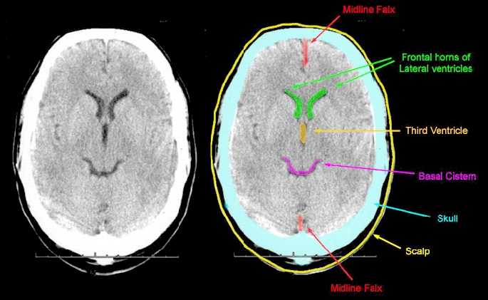
to normal appearances. The scan below is a slice through the human brain and you should imagine that you are viewing it as if looking up from the patient's feet. Therefore, the patient's left is to the right of the screen. The shape of the ventricles is quite distinctive and they are shown outlined in green and orange. The presence of the third ventricle in the midline is one of the first things to look for. If the third ventricle is either not visible, or shows signs of shift away from the midline, this suggests that there is an abnormality. The basal cisterns is the fluid filled space around the back of the midbrain outlined here in purple. Blood clots, or swelling of the brain may cause this to become narrowed, or not visible altogether. Note in this scan, that the frontal horns of the lateral ventricles are symmetrical, with the septum between them in the midline.


Back

Case 2 - Acute Subdural Haematoma Demonstrating Midline Shift:
Midline shift >5mm
Intracranial haematoma - non evacuated
Cortical contusion >1cm in diameter

Obliteration of 3rd Ventricle (not seen - refer to normal CT scan)

This CT scan shows a right sided acute haematoma, as well as an associated cerebral contusion (bruising). The true midline has been outlined by yellow dots and you can see that the frontal horns of the lateral ventricles have been pushed over to the left. In addition, the third ventricle is now not visible and it is also extremely difficult to make out the basal cisterns. This scan demonstrates four of the features which are included on the Early Outcome Form, namely midline shift greater than 5mm, intracranial haematoma - non evacuated, cortical contusion greater than 1cm in diameter and obliteration of the third ventricle. This haematoma requires surgical evacuation, otherwise deterioration of the patient's condition is inevitable.


Back

Case 3 - Acute Subdural Haematoma
Intracranial haematoma - non-evacuated

This scan demonstrates a left sided acute subdural haematoma. The scan is taken through a slightly higher part of the brain and shows the bodies of the lateral ventricles. The left lateral ventricle has been compressed and the midline is deviating to the right. The right lateral ventricle is actually slightly larger than normal and this is because the increased pressure is preventing escape of the cerebrospinal fluid from that ventricle. Dilatation of the contralateral ventricle like this indicates that there is very significant pressure on the brain. This scan would be classified as "Intracranial haematoma - non evacuated" on the Early Outcome Form.


Back

Case 4 - Acute Extradural Haematoma:
Intracranial haematoma - non-evacuated

This scan shows another intracranial haematoma, namely an extradural. You will note that this haematoma has a concave shape, a bit like the human lens and this is because it is occurring between the bone and the dura and is not actually lying on the surface of the brain itself. The points of attachment of the dura limit the extension of this haematoma anteriorly and posteriorly. You can see that there is shift of the midline. Look at the frontal horns in their relation to the falx cerebri (falx cerebri is outlined on the normal scan). This scan would be classified "Intracranial haematoma - non evacuated."


Back

Case 5 - Diffuse Axonal Injury:
One or more petechial haemorrhages within the brain

The presence of petechial haemorrhages is usually an indication of a very severe primary brain injury. Petechial haemorrhages tend to occur at the interface of grey and white matter. It can also occur in the dorsolateral quadrant of the midbrain at the middle orange arrow, as well as elsewhere within the brain substance. Note on this scan, that the lateral ventricles and the third ventricle are visible and there is no midline shift. It is often a characteristic of diffuse axonal injury, in which there are numerous petechial haemorrhages that there is no evidence of brain swelling, or midline shift. This scan would be classified as showing one, or more, petechial haemorrhages within the brain.


Back

Case 6 - Cerebral Contusion:
Cortical contusion >1cm in diameter

This is a scan of a patient who has sustained a severe head injury. There is extensive bruising of the right side of the brain, showing up as a large, diffuse grey area. You can also see that there are patches of white within the grey area. This represents bleeding. The grey area represents swelling (oedema). The area of the cortical contusion is outlined in purple. You will normally find a centimetre scale at the right hand side of a CT scan. This scan would be classified on the Early Outcome Form as "Cortical contusion - greater than 1cm in diameter.